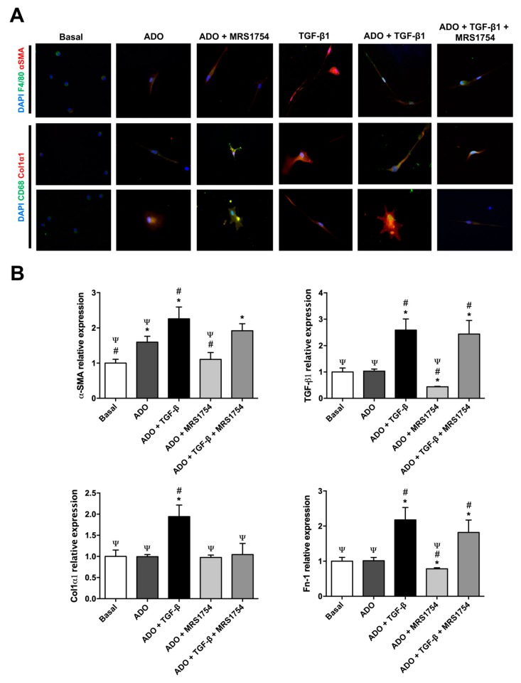
Figure 6.
In vitro blockade of A2BAR decreased myofibroblast marker expression in macrophages. (A) Immunocytofluorescence of macrophage (F4/80 and CD68; green) and myofibroblast (α-SMA and Col1α1; red) markers in human macrophages cultured in Roswell Park Memorial Institute (RPMI) high D-glucose medium 0.5% FBS (basal) treated with 1 µM adenosine (ADO), 10 ng/mL TGF-β1, and 10 nM MRS1754 for seven days. DAPI (blue) was used as a counterstain. Cells selected from the image captured with a 400× magnification. (B) mRNA expression of myofibroblast markers α-SMA, TGF-β1, Col1α2, and Fn-1 by RT-qPCR in human macrophages cultured under basal condition, adenosine (ADO), TGF-β1, and MRS1754 for seven days. HPRT mRNA expression was used for normalization. Graphs represent the mean ± S.D. * p < 0.05 versus basal; # p < 0.05 versus ADO; Ψ p < 0.05 versus ADO + TGF-β1. n = 3.

