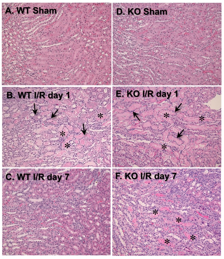
Figure 3.
Deletion of VDAC1 exacerbates ischemia-induced damage to morphology of renal proximal tubules and impairs renal tissue regeneration after acute injury. On days 1 and 7 after ischemia, kidneys were harvested, immersed, fixed in 4% neutral-buffered formaldehyde, and embedded in paraffin. Thin (4–8 μm) sections were cut and stained with hematoxylin-eosin. Renal histology of the cortical-medullary junction was assessed in kidney sections from wild type (WT, A–C) and VDAC1-deficient (KO, D–F) mice. The images represent kidneys from sham-operated mice (A,D) and mice sacrificed on day 1 (B,E) and day 7 (C,F) after ischemia. * Necrotic tubules. The arrows mark inflammatory cell rings accumulated around necrotic tubules. The images were captured using Nikon Eclipse E800 microscope and Nikon 10× Plan Apo objective. Magnification: 122×. The sections are representative of kidneys from three WT and four KO sham-operated mice at each time point after ischemia (I/R) in different experiments.

