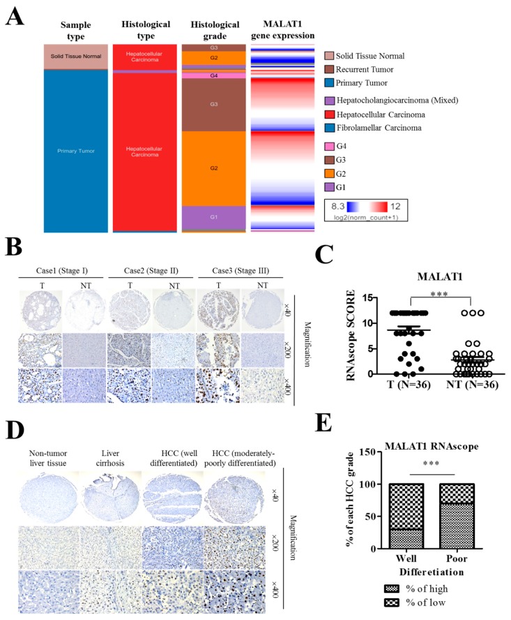
Figure 2.
LncRNA MALAT1 overexpression in liver cancer positively correlates with poor cellular differentiation status and disease progression. (A) In the TCGA liver cancer (LIHC) cohort (n = 438) significantly positive correlation between MALAT1 expression and cellular differentiation (histologic) grade was observed. G1, grade 1 = well differentiated; G2, grade 2 = moderately differentiated; G3, grade 3 = poorly differentiated; G4, grade 4 = undifferentiated/anaplastic. (B) Representative RNAscope images (T = Tumor; NT = Non-tumor) and (C) Boxplots graph depicting the manual scores showing positive correlation between increased expression of MALAT1 and HCC pathological stage. (D) Representative RNAscope images and (E) stacked histogram showing the differential expression of MALAT1 in non-tumor liver tissue, liver cirrhosis, well-differentiated HCC and moderately-poorly differentiated HCC tissues. *** p < 0.001.

