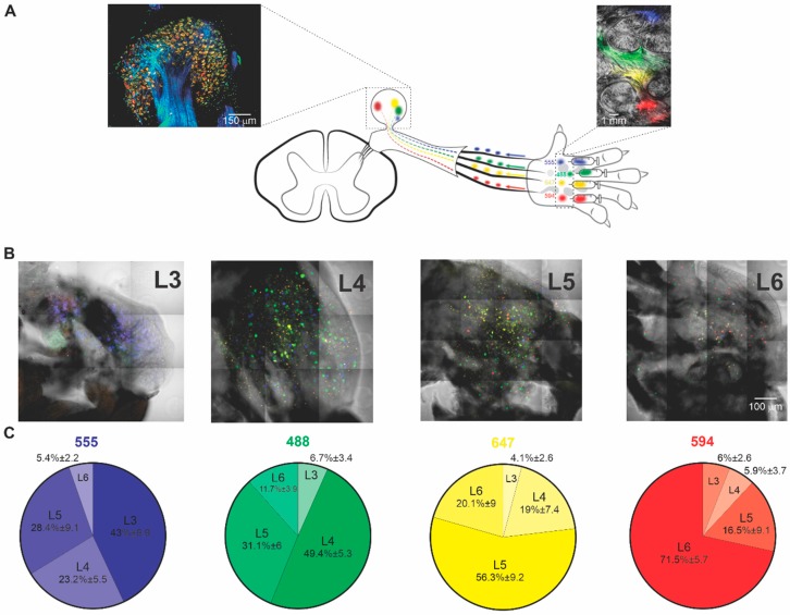
Figure 1.
Retrograde labeling of dorsal root ganglion (DRG) somata that innervate distinctive areas of the paw. (A) Scheme depicting the Painbow approach. Four spectrally different fluorophores AF 488—green, AF 555—blue, AF 594—red, AF 647—yellow, conjugated to wheat germ agglutinin (WGA), were injected into the paw in the illustrated order. The dyes were uptaken by the axonal terminals innervating the injected paw skin and transported to DRG somata. The labeling of the specific DRG reflects the area in the paw this DRG innervates. Left inset, a high-resolution confocal fluorescent image of L5 DRG excised 40 h after injecting the dyes into the skin. Right inset, the fluorescent image of the dissected paw skin obtained 40 h after the dye injection. Note that colors remained in the paw 40 h after the injection, allowing the correlation between the skin area and labeled DRG somata. Note differently labeled somata of DRG neurons. (B) Collaged fluorescent photomicrography of representative individual L3-L6 DRGs excised from naïve rats. Colored dots are DRG somata, retrogradely labeled from their innervation targets. (C) Distribution of injected dyes among lumbar DRGs of naïve rats. Note that as expected form the innervation patterns, AF555 injected into the medial part of the paw is mainly present in L3 DRG; AF488 injected laterally to AF555 is mainly present in L4 DRG; AF647 injected into the middle of the paw is mainly present in L5 DRG, and AF594 injected into the lateral paw is mainly present in L6 DRG. Data from five DRGs for each level from 5 rats, chi-square independence test (9) = 236.58, p < 0.001, suggesting that the DRG segments and the distribution of the colors are not independent of each other.

