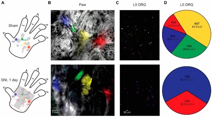
Figure 3.
The Painbow detects changes in innervation. (A) Scheme representing the injection arrangement. The four dyes were injected into the paw one day after either sham (upper panel) or SNI (lower panel) surgery. The dyes were injected to sham and SNI groups in a similar arrangement and in accordance with the denervation pattern (depicted in the lower panel, as a grey “T-shaped” area), such that AF555 and AF594 were injected into medial and lateral areas (respectively) of the paw that does not undergo denervation. AF488 and AF647 were injected into the areas which were denervated after SNI. (B) Representative fluorescent images of the skin demonstrating the arrangement of the injections one day after sham surgery (upper panel) and 1 day after SNI (lower panel). A representative of six rats for sham and SNI groups (C). Representative fluorescent images of L5 DRGs from sham-operated (upper panel) and SNI (lower panel) rats, removed 40 h after dyes were injected into the skin. Note that in DRG from a sham-operated animal, all colors are present, whereas in DRG from the animal after SNI, only colors injected into areas that are not underwent denervation (AF555 (blue) and AF594 (red)) are present. A representative of 6 rats for sham and SNI groups. (D) Distribution of dyes in L5 DRGs from sham-operated animals (upper panel) and rat after SNI (lower panel). Note that in L5 DRG from sham-operated rats the majority of the cells were labeled with AF647, but all colors were present, suggesting that L5 DRG neurons send their axons into all labeled skin areas shown in A. In animals after SNI no cells labeled with the dyes injected into the denervated areas were found, n = 6 rats in each group.

