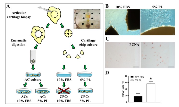
Figure 1.
Experimental design of cell cultures (articular chondrocytes (ACs) and chondro-progenitors (CPCs) from human articular cartilage biopsies. (A) Representative illustration of biopsy handling to obtain ACs culture and cartilage chip culture. (B) Optical images of cartilage chips after 15–20 days in culture with cells coming out to the medium supplemented with platelet lysate (PL) versus fetal bovine serum (FBS) and (C) representative immunohistological distribution of proliferating cell nuclear antigen (PCNA)-positive cells inside tissue in both culture conditions (N = 3). (D) Histogram showing the percentage of PCNA-positive cells in cartilage chips maintained in culture with FBS or PL. Data are represented as mean ± SEM (N = 3, * p < 0.05 versus ACs 10% FBS by Student’s t-test analysis). All scale bars correspond to 100 μm.

