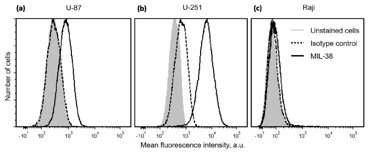
Figure 1.
Histograms demonstrating flow cytometry analysis of the binding of anti-GPC-1 antibody MIL-38 to GBM cells U-87 (a), U-251 (b), and lymphoma cells Raji (c). The light gray filled histogram represents unstained cells, dashed black line shows the cells incubated with an isotype control antibody, and black line shows the cells incubated with MIL-38.

