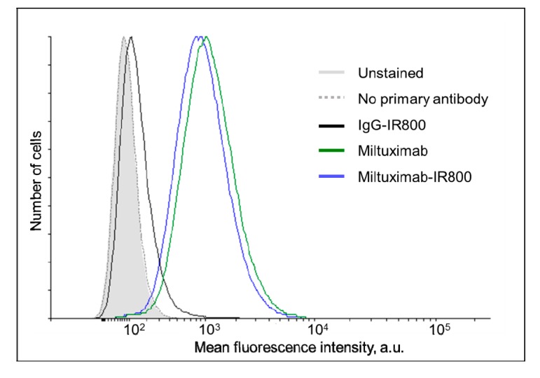
Figure 3.
Histograms demonstrating the flow cytometry analysis of the labelling of U-87 cells by Miltuximab®-IR800 (solid black line), IgG-IR800 (dark gray line), or unconjugated Miltuximab® (dotted black line). After incubation with the conjugates, the cells were stained by an anti-human-IgG antibody conjugated to a fluorescent dye AlexaFluor488. The light gray-filled histogram represents unstained cells and the dotted gray line shows the cells incubated only with the secondary antibody.

