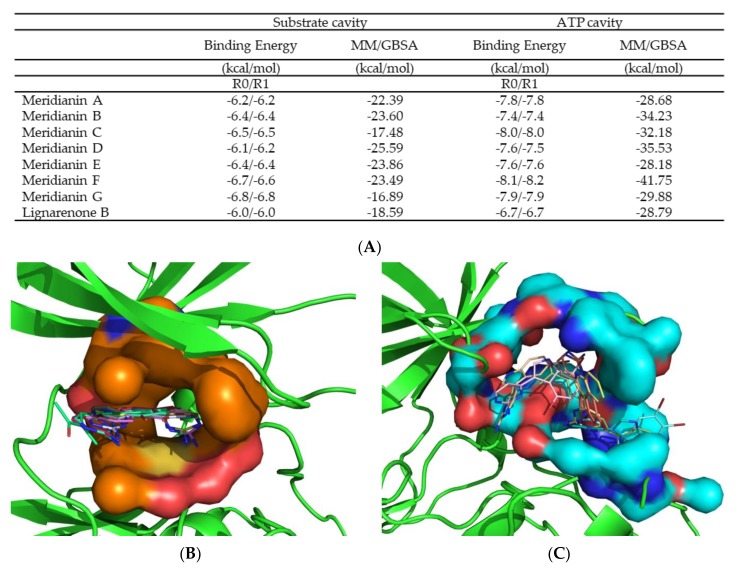
Figure 4.
Representation of the two binding cavities ATP and substrate in surface, the binding mode of the marine molecules and the summary of the binding energy results. (A) Binding energy results after docking calculations and after 1 ns molecular dynamics (MD) simulations with molecular mechanics/generalised born surface area MM/GBSA calculations. All energies values are in kcal/mol. (B) The ATP pocket with all the meridianins and lignarenone B. (C) The substrate pocket also with all the meridianins and lignarenone B. Both images represent the last frame after MD simulation. Meridianin A-G colours: Peach, blue, tan, orange, pink, cyan and yellow. Lignarenone colour: green.

