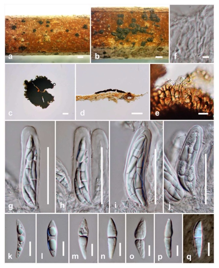
Figure 3.
Pseudopalawania siamensis (holotype) (a,b) Appearance of ascomata on substrate. (c) Squash mounts showing ascomata. (d) Section of ascoma. (e) Peridium. (f) Pseudoparaphyses. (g–j) asci. (k–p) Ascospores. (q) Ascospores in Indian ink. Scale bars: a, b = 500 µm, c, d = 100 µm, g–j = 50 µm, e, k–q = 10 µm, f = 5 µm.

