Figure 2.
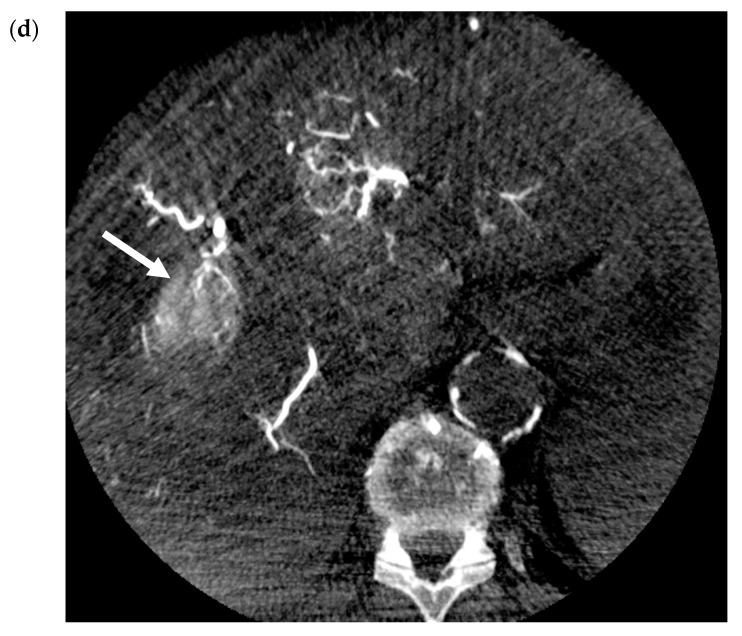
Transarterial Embolization (TAE) of HCC. (a) Pre-procedure triphasic contrast-enhanced CT in the arterial phase. Mass with arterial hyperenhancement in segment 8 of the right lobe of liver (white arrow), consistent with hepatocellular carcinoma (HCC). (b) Digital subtraction angiogram of the right hepatic artery pre-bland embolization. Mass (white arrow) is supplied by the segment 8 branch of the right hepatic artery (black arrow). (c) Digital subtraction angiogram of the right hepatic artery post-bland embolization of the segment 8 branch of the right hepatic artery. A combination of 40-120-µm microspheres and 100-µm polyvinyl alcohol (PVA) particles were used. Angiogram demonstrates the stasis of iodinated contrast in the segment 8 arterial branch with pruning of the distal branches and no opacification of the mass. (d) Intra-procedural cone-beam CT immediately post-bland embolization. Retention of iodinated contrast and particles in the treated tumor (white arrow) identical to pre-procedure post-contrast images indicative of complete tumor coverage.


