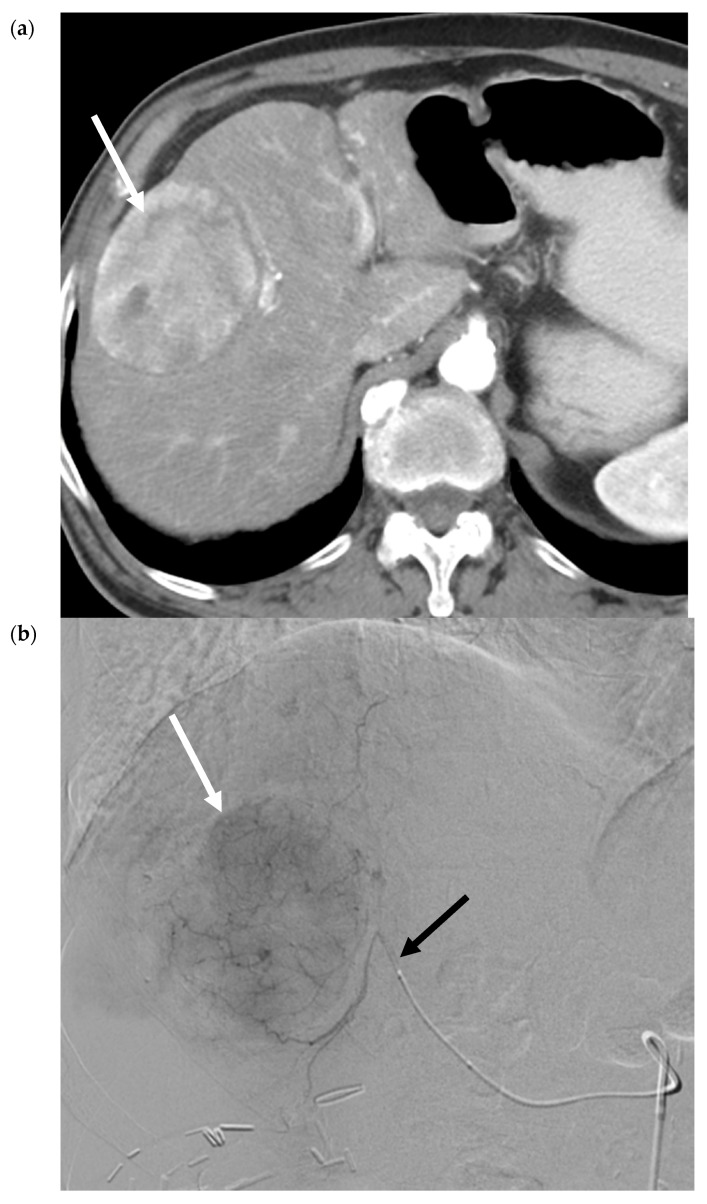
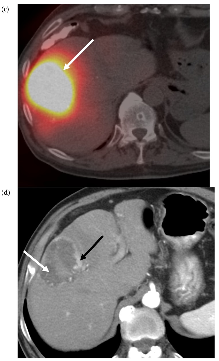
Figure 4.
90Y Radiation Segmentectomy for Solitary HCC. (a) Pre-procedure triphasic contrast-enhanced CT in the hepatic arterial phase. Mass (white arrow) with arterial hyperenhancement in the anterior sector of the right lobe of the liver, consistent with solitary HCC. (b). Digital subtraction angiogram of the anterior division of the right hepatic artery prior to administration of glass 90Y microspheres. Mass (white arrow) is supplied by the anterior division of the right hepatic artery (black arrow). (c) SPECT image immediately post-90Y radioembolization. High activity of 90Y microspheres in the liver tumor (white arrow) is demonstrated without evidence of nontarget delivery. (d) Post-procedure triphasic contrast-enhanced CT in the arterial phase performed 6 weeks post-treatment. Marked decrease in arterial hyperenhancement of the anterior right hepatic tumor. Overall, the mass is hypoenhancing (white arrow) due to the treatment effect, with a rim and few areas of residual enhancement (black arrow).


