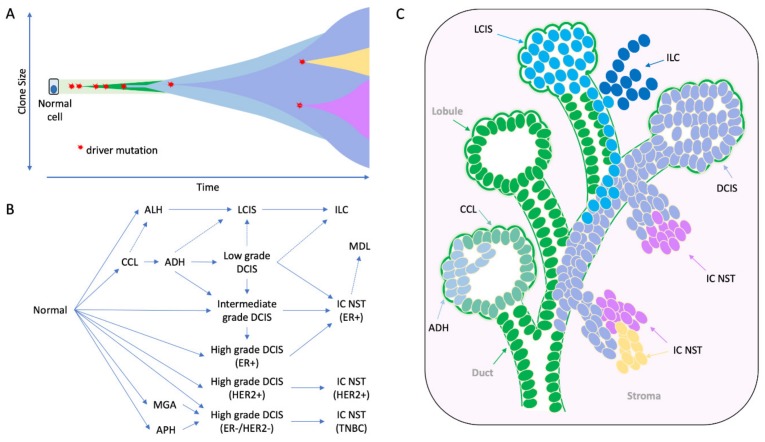
Figure 2.
The morphological and molecular evolution of breast cancer. (A) Hypothetical schematic showing how the mutation of cancer genes drives the clonal and subclonal evolution of cancer (adapted from [55]). Key early driver genes impact the subsequent lineage and tumour type that arises, including mutations in PI3KCA in ER+ tumours, CDH1 in lobular lineage, TP53 in high grade ER- tumours, ETV6-NTRK3 and MYB-NFIB translocations in secretory and adenoid cystic carcinomas respectively. (B) the multistep model of breast cancer showing morphological stages of development from normal epithelium. This simplified model is based on the evolution of ER positive and ER negative breast cancer, as portrayed in more detail elsewhere [49,50,51]; evidence derived from morphological evaluation and the frequency with which lesions are co-localized, as well as molecular evidence showing co-localized lesions share identical mutations indicating clonal relatedness. (C) Cartoon to illustrate how this might arise in a ‘sick lobe’, that is a clonal outgrowth of apparently morphologically normal-looking epithelial cells (green), which harbour early genetic changes. In some areas of the lobe, the earliest morphologically abnormal changes may appear in some terminal duct-lobular units (lobule) as columnar cell lesions. These lesions are considered precursors of ADH (light blue cells) and DCIS (purple cells), which arise in lobules and may travel down ducts. The mutation or loss of CDH1 (E-cadherin) triggers the evolution of the ’lobular lineage’ (sky blue cells) as ALH then LCIS (lobular neoplasia); these cells may travel down ducts underneath the normal epithelial lining (pageotoid spread). Both LCIS and DCIS are genetically advanced lesions and so likely exhibit sub-clonal mutations. As these neoplastic cells can travel along ductal structures then this means invasion can occur at multiple sites giving rise to multifocal invasive disease (ILC, IC NST), which continues to undergo subclonal change. CCL: columnar cell lesion; ADH: atypical ductal hyperplasia. ALH: atypical lobular hyperplasia, APH: atypical apocrine hyperplasia; DCIS: ductal carcinoma in situ; LCIS: lobular carcinoma in situ; IC NST: invasive carcinoma no special type; ILC: invasive lobular carcinoma; MDL: mixed ductal lobular carcinoma; MGA: microglandular adenosis.

