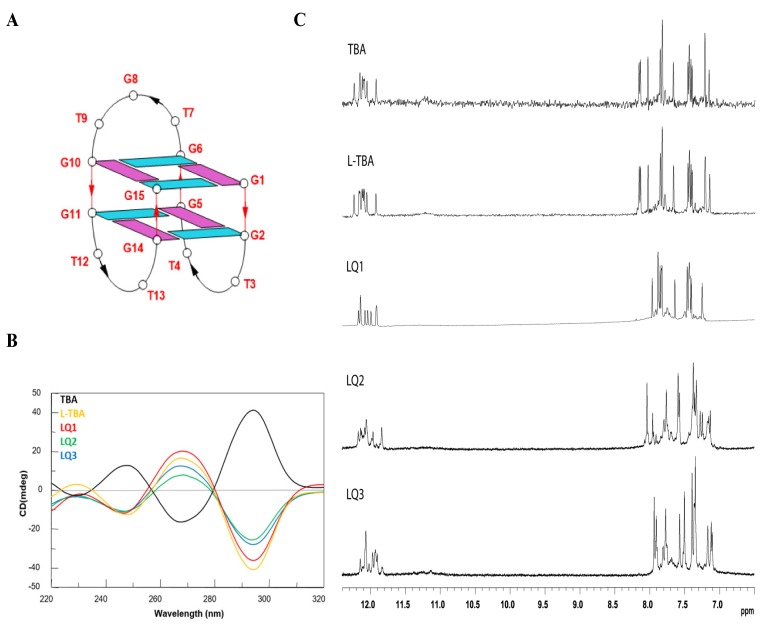
Figure 1.
Structural investigations of the thrombin binding aptamer (TBA) derivatives. (A) Schematic representation of the G-quadruplex structure adopted by TBA. Guanosines in syn and anti glycosidic conformations are in purple and light blue, respectively. (B) CD spectra at 20 °C of the modified TBAs and their natural counterpart at 50 µM ODN strand concentration in a buffer solution 10 mM KH2PO4/K2HPO4, 70 mM KCl (pH 7.0). (C) Aromatic and imino proton regions of the 1H NMR spectra (500 MHz) of the ODNs investigated (Table 1). See the Materials and Methods section for details.

