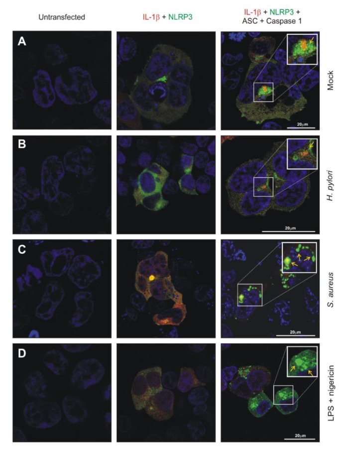
Figure 6.
Confocal laser scanning microscopy was used to analyze the reconstructed HEK293-NLRP3-INSOME cells in mock control (A), H. pylori-infected (B), S. aureus-infected (C) and E. coli LPS/Nigericin-treated (D). EGFP-NLRP3 and mCherry-pro-IL-1β were visualized using their characteristic colors and NLRP3 inflammasome in various conditions mentioned above were marked with yellow arrows. The respective column heading shows the transfection status of each cell before or after infection or treatment.

