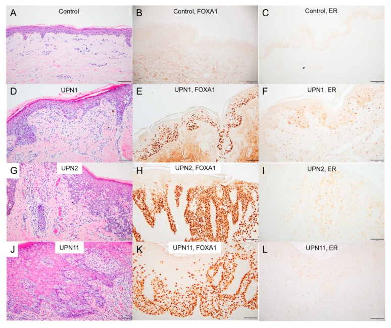
Figure 4.
Immunohistochemical analyses of EMPD lesions and normal epidermis. Skin specimens from a healthy donor (A–C); UPN1, the case with the GAS6-FOXA1 fusion (D–F); UPN2, the case with the FOXA1 promotor mutation (G–I); and UPN11, a case with no detectable mutation (J–L) stained with anti-FOXA1 (B,E,H,K) and anti-estrogen receptor alpha (C,F,I,L) antibodies (scale bars, 100 μm).

