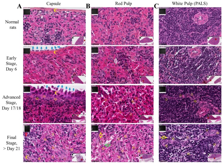
Figure 5.
Spleen colonization by M5-T1 cells. High magnification views of the successive stages (×800). Scale bars represent 25 µm, inserts showing general views (×25). (A) Invasion of the capsule (C, in yellow). The open white arrow shows clustering of reticular cells. Open yellow arrows indicate M5-T1 tumor cells crossing the capsule or present in the red pulp. Vicinal tumor, T in red. (B) Red pulp. Early stage: the architecture of the red pulp is preserved, large open white arrow indicating clusters of lymphoid cells on the tumor side attached to the spleen surface. Advanced stage and onwards: open yellow arrows indicate numerous tumor cells actively moving inside the red pulp. Final stage: the large green arrow shows empty spaces, attesting a decrease in the density of normal cells. The large yellow arrow indicates clusters of lymphoid and tumor cells. (C) White pulp, periarteriolar lymphoid sheath (PALS). Early stage: the architecture of the follicle is preserved on this cross-section of the central artery. Advanced stage: open yellow arrows indicate tumor cells penetrating the PALS on this longitudinal section of the central artery. Final stage: tumor cells present everywhere following the destruction of the stromal framework of reticular fibers.

