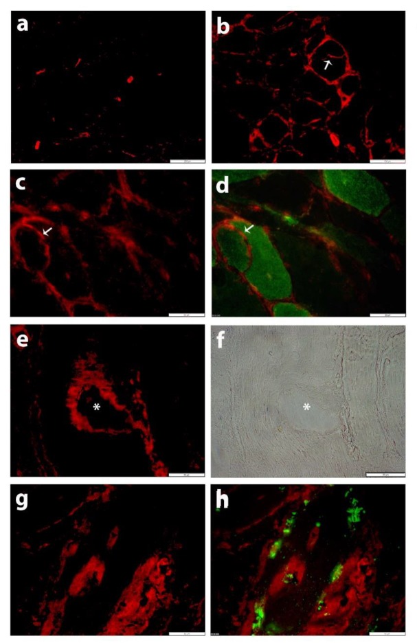
Figure 1.
Immunofluorescent staining for SLC5A3 (CY3, red) with antibody Ab1. (a) In normal skeletal muscle tissue, staining is restricted to blood vessels and occasional partial staining of muscle fiber membranes. (b–h) SLC5A3 staining (CY3, red) is increased in the muscle tissue from patient IBM7: (b) SLC5A3 staining is observed on blood vessels, endomysial inflammatory cells and the muscle fiber membrane, including the membrane of a splitting fiber (arrow), which is a pathological feature associated with muscle tissue damage and regeneration. (c,d) Discontinuous membranous SLC5A3 staining is present on a small fiber that is CD56+ (AlexaFluor488, green, arrow). (e) SLC5A3 staining is accentuated on the rims of a vacuole, as confirmed by the phase contrast image (f, asterisk). (g,h) SLC5A3 staining associates with the presence of inclusions, visualized as sequestosome 1/p62-positive (AlexaFluor488, green). Scale bars 200 µm (a), 100 µm (b), 50 µm (c–h).

