Figure 1.
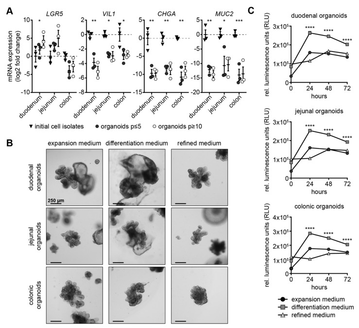
Establishment of new culture conditions for canine intestinal organoids. (A) RT-qPCR analysis of initial cell isolates (triangles), organoids with up to five passages (dark circles) and organoids with 10 or more passages (light circles) isolated from duodenum, jejunum or colon using LGR5, VIL1, CHGA and MUC2 primer. log2 fold changes normalized to expression of initial cell isolates are shown with scatter dot plots; mean is shown; whiskers present SEM; * p < 0.05, ** p < 0.01 and *** p < 0.001, statistical analysis given in detail in Supplementary Tables S2–S4; n = 3 individual dogs. (B) Light microscopic images of organoids derived from duodenum, jejunum and colon cultivated in expansion, differentiation and refined medium four days after seeding; scale bars represent 250 µm. (C) Mean activity of Annexin V-induced luciferase is shown for 0, 24, 48 and 72 h after addition of substrate to duodenal, jejunal and colonic organoids in expansion (circle), differentiation (square) and refined medium (triangle); whiskers represent SEM; **** p < 0.0001; 50 organoids seeded per replicate; n = 6 replicates.

