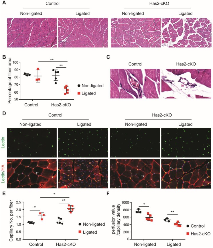
Figure 2.
Loss of hyaluronan in ECs leads to muscle damage and excessive angiogenesis after femoral artery ligation. (A) Representative images of H&E staining and (B) quantification of myocyte area in ligated and non-ligated adductor muscle of Has2-cKO (n = 5) and control (n = 3) mice (scale bar = 100 µm). (C) Representative images of H&E staining show leaky vessels in Has2-cKO mice (n = 5) after single femoral artery ligation (scale bar = 20 µm). (D) Representative images of BS1-lectin staining for ECs localization (green) and HA staining with the HA binding probe Ncan-dsRed (red). HA staining is used for the myofibroblasts visualization and are found to be surrounded by HA staining as for example depicted by the white arrows. (E) quantification of capillary densities in ligated and non-ligated gastrocnemius muscle of Has2-cKO (n = 5) and control (n = 3) mice. The capillary density is defined as capillary number per myofibroblast. (F) quantification of capillary perfusion capacity, calculated from measured LDPI perfusion value, normalized by capillary density in Has2-cKO (n = 5) and control (n = 3) mice after fully recovery. Values are given as mean ± SD. * p < 0.05, ** p < 0.01.

