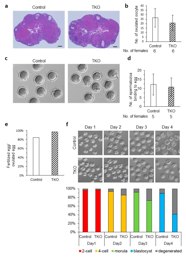
Figure 4.
Phenotypic analysis of Oosp family KO females. (a) Histological analysis of ovaries in Oosp heterozygous KO (Control) and homozygous KO (TKO) mice. Scale bars = 200 µm. (b) Average number of oocytes ovulated by a hormone primed female. Four-week-old females were examined (n = 6). (c) Sperm ZP-binding assay. Spermatozoa were inseminated with control and KO oocytes from which cumulus were removed. (d) Average number of spermatozoa bound to ZP. Three-week-old to 14-week-old females were used. (n = 5) (e) Average fertilizing ratio: 133 heterozygous and 106 TKO oocytes were examined. (n = 5) (f) Developmental ratio of embryos after in vitro fertilization. Superovulated oocytes from control and TKO females were examined. Two-cell embryos, four-cell embryos, morulas, and blastocysts were observed at 24 h (Day1), 48 h (Day2), 72 h (Day3) and 96 h (Day4) after insemination. One hundred twelve and 103 eggs ovulated by four-week-old to 10-week-old heterozygous and TKO females were examined, respectively (n = 5, p < 0.05).

