Figure 5.
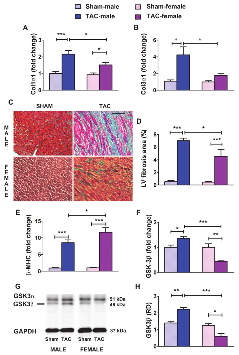
Regulation by pressure overload of relevant LV remodeling related elements in mice. A and B: Fold change in mRNA levels of Col1α1 (A) and Col1α3 (B) in the LV from male and female mice subjected to sham surgery or transverse aortic constriction (TAC) for 4 weeks (sham males, n = 9; sham females, n = 11; TAC males, n = 15; TAC females, n = 12). (C) Representative images of LV sections showing myocardium stained with Masson’s trichrome. With this technique, muscle fibers are stained red, fibrosis is stained blue, the cytoplasm is stained light red or pink, and cell nuclei are stained dark brown to black. (D) Average fractional LV area of fibrosis in 4 sections from three to six mice per group. (E,F) Fold change in mRNA levels of β-MHC (E) and GSK-3β (F) in the LV from sham and TAC mice of both sexes. (G) Representative western blot images showing the protein levels of GSK-3β in the LV. (H) Average relative optical density (RD vs. GAPDH) quantified in three mice per group in two different experiments. Data are means ± SEM. * p < 0.05, ** p < 0.01; *** p < 0.001 (ANOVA followed by the Bonferroni post-hoc test).

