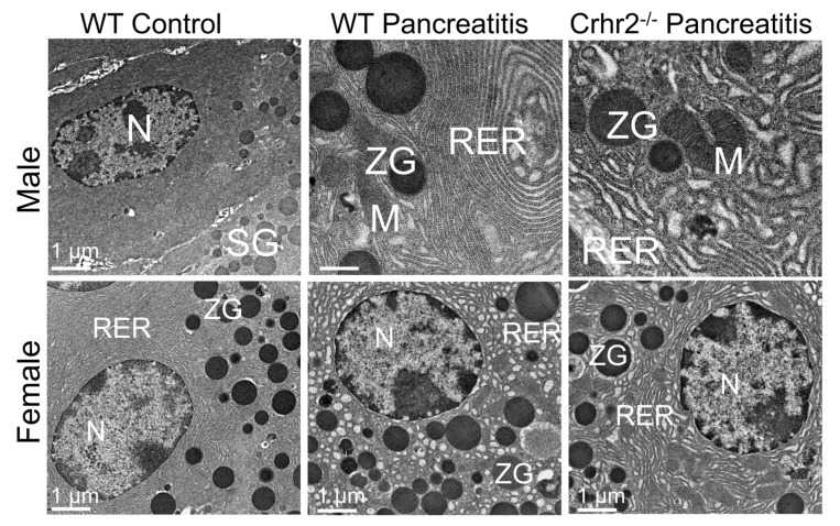
Figure 5.
Electron micrographs showing representative pancreatic acinar cell sections from control and pancreatitis male and female mice. Endoplasmic reticulum (RER) shows whorling and the distortion of cisternae; mitochondrial (M) swelling is also evident. N: nucleus; SG: secretory granules; WT: wild-type; ZG: zymogen granules. Scale bar: 1 µM. Modified from Kubat et al. [93].

