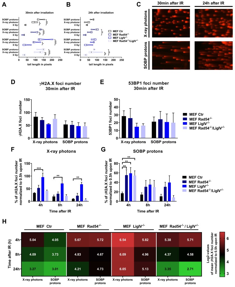
Figure 3.
DNA damage repair induced by X-ray photons or SOBP protons in MEFs. (A,B) Quantification of alkaline comet assay in wild type (Ctr), Lig IV-deficient (Lig IV−/−), Rad54-deficient (Rad54−/−) or Lig IV/Rad54 double-deficient MEFs (Rad54−/−/Lig IV−/−) at 30 min (A) and 24 h (B) after irradiation with 3 Gy of X-ray photons or SOBP protons. (C) Exemplary pictures of alkaline Comet assay of wild type MEFs (MEF ctr) 30 min or 24 h after X-ray photon or SOBP proton irradiation. (D) γH2A.X and (E) 53BP1 foci number 30 min after irradiation with 3 Gy of X-ray photons and SOBP protons. (F,G) Quantification of DNA repair kinetics of MEFs harboring deficiencies in Rad54, Lig IV or Rad54 and Lig IV determined by quantification of γH2A.X foci. Data represent values normalized to 30 min after irradiation with 3 Gy. (H) Comparison of radiation source effects in tested cell lines as log2 values of γH2A.X foci quantification. Data represent mean SEM from three independent experiments. Two-way ANOVA with Tukey’s multiple comparisons post-test; * p < 0.05; ** p < 0.01; *** p < 0.001; **** p < 0.0001.

