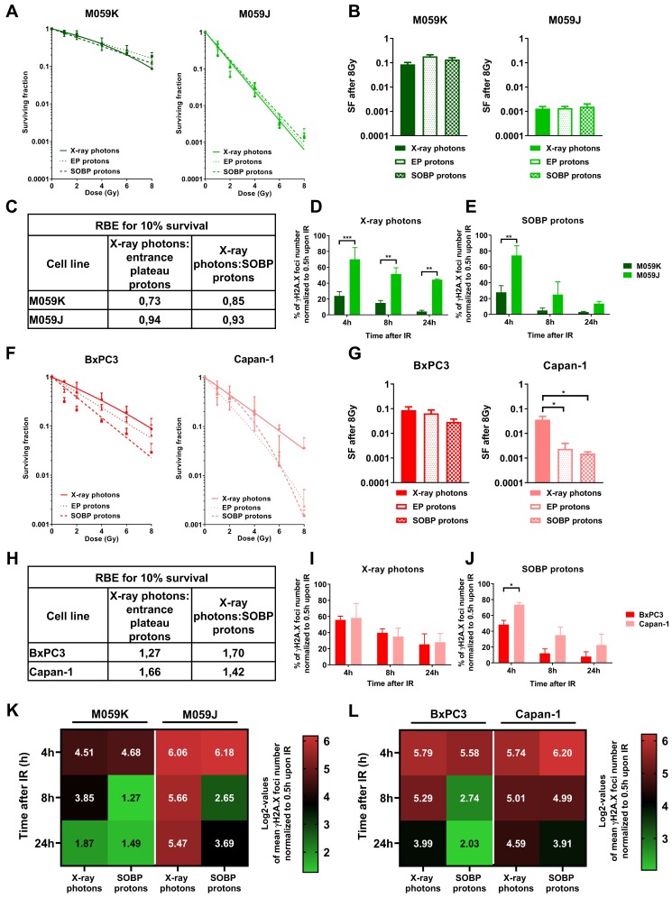
Figure 4.
Effects of X-ray photons, EP and SOBP protons on clonogenic cell survival and kinetics in formation and removal of γH2A.X foci in cancer cells. (A,B) Clonogenic survival assay of DNA-PKcs-deficient M059J and DNA-PKcs-proficient M059K glioblastoma cells irradiated with X-ray photons, EP and SOBP protons (1–8 Gy). (C) RBE values calculated for 10% of clonogenic cell survival of the indicated cell lines. (D,E) γH2A.X foci removal over time (4–24 h) normalized to initial foci number at 30 min in M059J and M059K cells upon irradiation with 3 Gy of X-ray photons (D) and SOBP protons (E). (F,G) Clonogenic survival assay of pancreatic cancer cells with BRCA2-deficiency (Capan-1) and BRCA2-proficiency (BxPC3). (H) RBE values calculated for 10% of clonogenic cell survival of the indicated cell lines. (I,J) γH2A.X foci removal over time (4–24 h) normalized to initial foci number at 30 min in Capan-1 and BxPC3 cells upon irradiation with 3 Gy of X-ray photons (I) and SOBP protons (J). (K,L) Comparison of radiation quality effects in tested cell lines as log2 values of γH2A.X foci quantification. Data represent mean values ± SD (A,F) or ± SEM (B,D,E,G,I,J) from three independent experiments. One-way ANOVA with Tukey’s multiple comparisons post-test (B,G) or two-way ANOVA with Tukey’s multiple comparisons post-test (D,E,I,J); * p < 0.05; ** p < 0.01; *** p < 0.001.

