Figure 4.
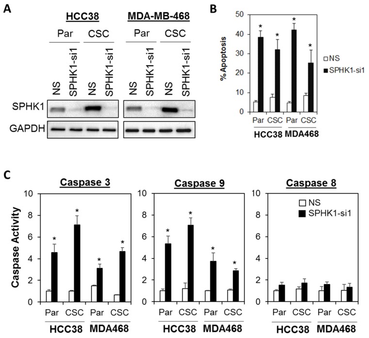
Depletion of SPHK1 induces cell apoptosis in breast CSC and non-CSC cultures. (A) Efficient depletion of SPHK1 expression was achieved using a single lentiviral shRNA construct in HCC38 and MDA-MB-468 adherent parental (Par) and CSC cultures. Non-silencing (NS) controls were included for accurate assessments of knockdown efficiency; GAPDH was detected as a loading control. (B) The proportion of cells undergoing apoptosis was assessed by Annexin V/7-AAD flow cytometry at 72 h following SPHK1 knockdown. Note the significant induction of apoptosis in transduced parental and CSCs. (C) Depletion of SPHK1 expression activates caspase 3/7 and 9, but not caspase 8. Both parental and CSCs were transduced with lentiviral shRNA targeting endogenous SPHK1. Caspase activities were determined using the CaspaseGlo assay at 72 h after transduction. Bars represent the means ± s.d. of three independent experiments. Asterisks (*) indicate statistical significance compared with non-silencing (NS) control cells (p < 0.01, Student’s t-test).

