Figure 2.
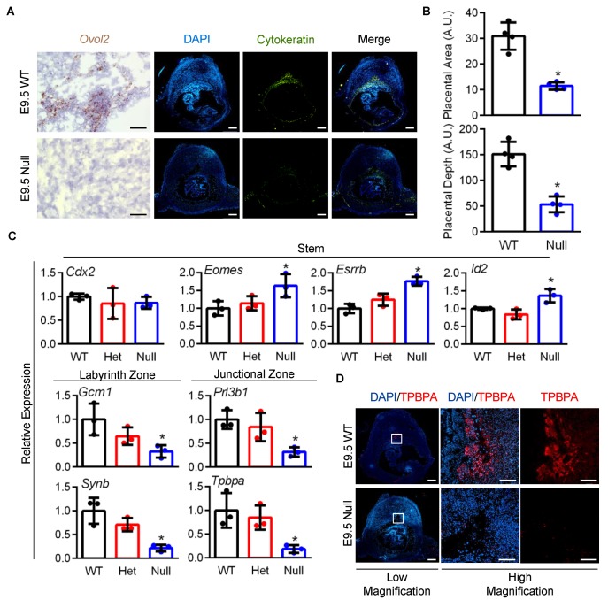
Placental development is impaired in Ovol2-deficient embryos. (A) Evaluation of Ovol2 expression in wild-type (WT) and Ovol2-null placentas on E9.5. Immunohistochemical staining of cytokeratin was used to denote the location of placentas. Note poor placental development in Ovol2-null placentas. (B) Quantitative analysis of placental area and depth comparing WT and Ovol2-null placentas. (C) Transcript analysis of stem (Cdx2, Eomes, Esrrb, and Id2) and differentiation markers (Gcm1, Synb, Prl3b1, and Tpbpa) comparing WT, heterozygote (Het), and Ovol2-null placentas at E9.5. (D) Immunohistochemical staining of differentiation marker TPBPA in WT and Ovol2-null placentas. Values significantly different from WT (N = 3–4 from different dams, P < 0.05) are denoted with an asterisk (*). Scale bars = 500 µm (low magnification) and 100 µm (high magnification). Graphs represent means (SD).

