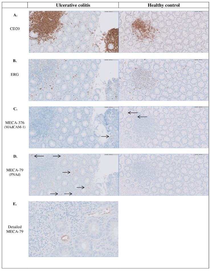
Figure 1.
Immunohistochemical staining indicating the presence of follicles and PNAd+ and MAdCAM-1+ venules in inflamed colonic tissue of UC patients and non-inflamed colonic tissue of a healthy control. Representative photomicrographs, with a magnification of x 20, of a colonic biopsy from a UC patient with (A) CD20 staining indicating B-cells (and follicles), (B) ERG staining indicating all venules, (C) MECA-367 staining indicating MAdCAM-1+ venules (pointed out with black arrows), (D) MECA-76 staining indicating PNAd+ venules (pointed out with black arrows) and (E) MECA-76 staining in detail with a magnification of x 40. Almost all PNAd+ and MAdCAM-1+ venules are located extrafollicular. MAdCAM-1, mucosal vascular addressin cell adhesion molecule-1; PNAd, peripheral node addressin; UC, Ulcerative colitis.

