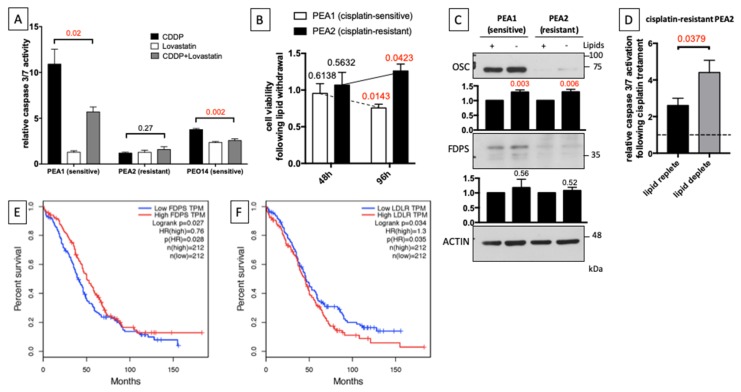
Figure 5.
Cholesterol synthesis inhibition increases chemoresistance in platinum-sensitive OC cells. (A) PEA1, PEA2, and PEO14 cells were treated with 5 μM Lovastatin and then treated with 20 μM cisplatin for an additional 48 h (when medium was changed, Lovastatin was added again). Subsequently, apoptosis was measured by a luminescent caspase 3/7 activity assay. Data are expressed as mean ± S.E.M. from four (PEA1 and PEA2) or six (PEO14) independent experiments with technical triplicates each. The numbers above the bars indicate the statistical significance (p-value), based on the two-tailed unpaired Student’s t-test (significant values highlighted in red). (B) Twenty-four hours after seeding PEA1 and PEA2 cells in standard culture medium, cells were cultured for additional 48 or 96 h in medium supplemented with lipid-stripped serum. Finally, cell viability was measured by an MTT-based cell viability assay. (C) Immunoblot of total lysates from PEA1 and PEA2 cells 48 h after lipid withdrawal from culture medium. Images are representative of five independent experiments. Bar plots below panels show mean ± S.E.M. of relative densitometric band intensity calculated by assuming each band of the control condition (normalized on Actin) = 1 (n = 5). The numbers above the bars represent the statistical significance (p-value) based on the Student’s t-test (significant values highlighted in red). (D) PEA2 platinum-resistant cells were cultured for 48 h in medium supplemented with lipid-stripped serum and then treated with 40 μM cisplatin for additional 48 h. Apoptosis was measured by a luminescent caspase 3/7 activity assay. Data are expressed as mean ± S.E.M. from four independent experiments with technical triplicates each. The numbers above the bars indicate the statistical significance (p-value), based on the two-tailed unpaired Student’s t-test. (E,F) Kaplan–Meier estimates of the impact of FDPS (E) and LDLR (F) on overall survival in OC, according to The Cancer Genome Atlas (TCGA) database.

