Figure 7.
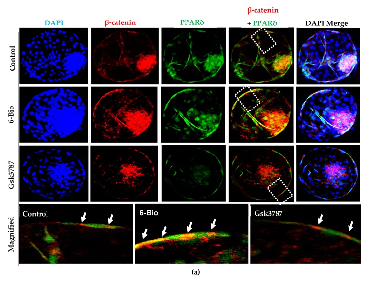
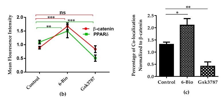
PPARδ co-localizes with β-catenin upon Wnt stimulation to regulate bovine embryonic development. (a) Immunofluorescence imaging showing co-localization between PPARδ (green) and β-catenin (red) is strengthened upon Wnt stimulation. Arrows indicate overlapping of PPARδ with β-catenin expression merge in (yellow). Nuclei counterstained with DAPI (blue). Higher magnification of the box regions shown at the bottom (b,c). Bar graph represents the quantification of mean fluorescence intensities and percentage of co-localization of PPARδ with β-catenin. All data are mean ± SEM from three individual sets of experiments (n = 20 BLs). * p < 0.05; ** p < 0.01; *** p < 0.001 indicates significant difference. Original magnification 100×.


