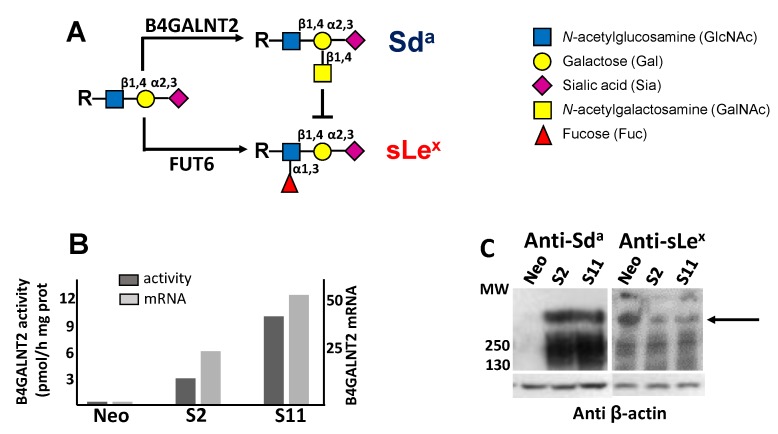
Figure 1.
Biochemical characterization of B4GALNT2-transfected cell lines. (A) the Sda and the sLex antigens derive from alternative and mutually exclusive terminations of a common α2,3-sialylated type 2 structure. (B) both the enzymatic activity (dark gray) and the mRNA (light gray) of B4GALNT2 were negligible in Neo cells, but strongly expressed in clones S2 and S11 as detected by RT-PCR and normalized with β-actin. (C) Western blot analysis of Neo cells and of B4GALNT2-transfected clones with anti-Sda (left) and anti-sLex (right) antibodies, revealing a partial replacement of the sLex antigen with the Sda (arrow).

