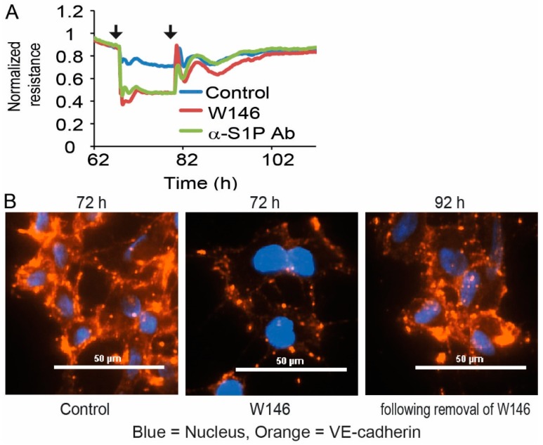
Figure 3.
Dependence and reversibility of EC barrier stability in HUVEC and EA.hy926. (A) Resistance following treatment with 3 µM S1PR1 antagonist W146 or 120 µg/mL of anti-S1P antibody Sphingomab, followed by the removal of the added substances. Line plot represents one experiment out of three with black arrows indicating the addition and removal of W146 or Sphingomab at the corresponding time points. (B) Immunofluorescence staining of VE-cadherin in HUVEC after addition of 3 µM S1PR1 antagonist W146, followed by removal of the added substance. Representative images from one out of three individual experiments are shown. Pictures were taken 6 h after addition of W146 and 12 h following removal of W146.

