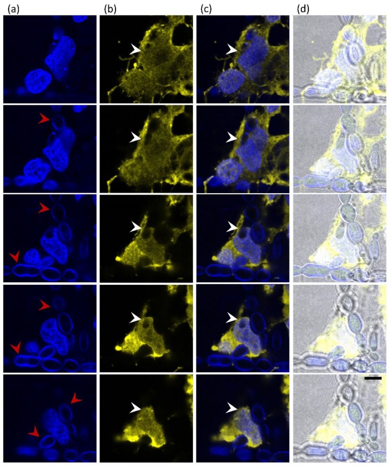
Figure 7.
Internalization of Exophiala dermatitidis into neuroblastoma cells SH-SY5Y after 48 h incubation. Five confocal images, imaged as Z-stack (top to bottom row), show the interaction of SH-SY5Y neuroblastoma cells with E. dermatitidis. (a) Fungi were detected with Calcofluor White (CFW) and SH-SY5Y nuclei with DAPI, both in blue. Red arrows point to pseudohyphae. (b) SH-SY5Y cytoplasm was stained in yellow using anti-GAPDH antibodies and secondary antibodies conjugated to Alexa 555. Penetrating fungal cells in the cytoplasm of neuroblastoma cells are clearly visible in the sequential cell sections (white arrows). (c) Merged images from images (a) and (b), (d) merged images including bright field (BF). Scale bar: 5 μm.

