Figure 1.
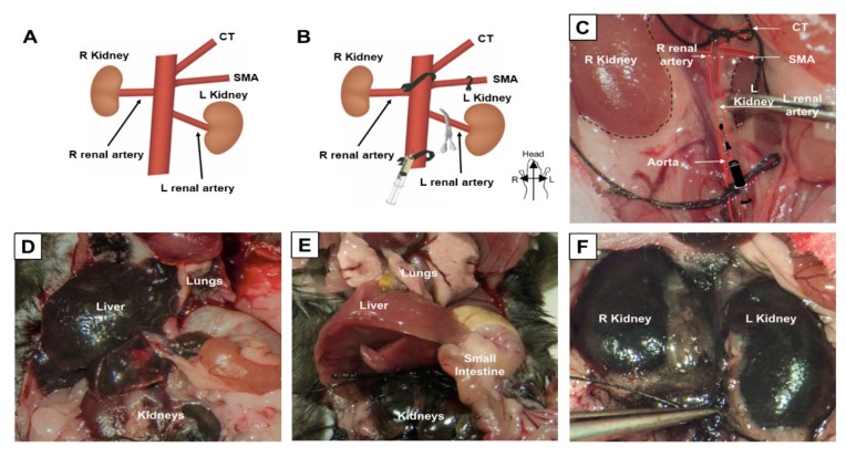
Selective ligation allows for selective kidney labeling. (A) Normal anatomy of the mouse abdominal aorta, showing origin of the celiac trunk (CT), superior mesenteric artery (SMA), renal arteries, and kidneys. (B) Suture ligation sites, including the proximal aorta between the origins of the CT and SMA, the SMA, and the distal aorta. A metal clip is placed temporarily on the left renal artery to allow delivery of therapeutics first to the right renal artery. (C) Selective ligation and clip placement shown inside the mouse abdomen. (D) Result of tattoo dye injection into the distal aorta without described ligation technique. (E,F) Result of tattoo dye injection with our ligation and clipping technique.

