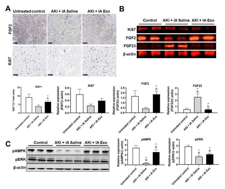
Figure 4.
Proliferation and regeneration markers. (A) Immunohistochemistry (IHC) of FGF2 and Ki67 in kidney tissue, and quantification of Ki67+ cells. (B) Western blot and quantification of Ki67, FGF2, FGF23, and β-actin from kidney lysate. (C) Western blot and quantification of pAMPK, pERK, and β-actin from kidney lysate. Measurements were taken at day 12. Each group has n = 3 pooled mice. Significant difference a p < 0.05: relative to untreated control group; b p < 0.05: relative to AKI + IA saline group.

