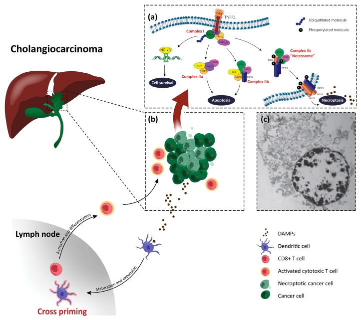
Figure 1.
A schematic overview of necroptosis in cholangiocarcinoma (CCA). (a) A simplified illustration of the intracellular pathways involved in cell fate in a necroptotic CCA cell. As reported in the text, tumor necrosis factor α (TNFα) activates its receptor, TNFα receptor 1 (TNFR1), which binds a series of proteins to form complex I. The ubiquitylation of receptor-interacting serine/threonine-protein kinase 1 (RIPK1) leads to cell survival, through the nuclear factor-kappa B (NF-κB) pathway. When this process is impeded, and caspase-8 is active, complex IIa will assemble, leading to RIPK1-independent apoptosis. The deubiquitylation of RIPK1 in the presence of activated caspase-8 leads to the assembly of complex IIb and, subsequently, to RIPK1-dependent apoptosis. The inhibition of caspase-8 leads to RIPK1/receptor-interacting serine/threonine-protein kinase 3 (RIPK3)/mixed lineage kinase domain-like (MLKL) interaction, forming complex IIc, also named the necrosome. Phosphorylated MLKL and RIPK3 translocate to the plasma membrane, opening membrane pores, and resulting in damage-associated molecular patterns (DAMPs) release. (b) The immunogenic response to DAMPs. Dendritic cells, activated by DAMPs, travel to lymph nodes, mature and expand, and present a tumor antigens to naïve CD8+ T cells, in a process called cross-priming. T cells are then activated and differentiate into cytotoxic T cells primed to specifically attack tumor cells. (c) The morphology of a necroptotic cell. Translucent cytoplasm, the swelling of organelles, patches of condensed chromatin within the nucleus, an increased cell volume, and a disrupted cell membrane can be visualized by electron microscopy. Picture adopted from Vandenabeele et al. [20].

