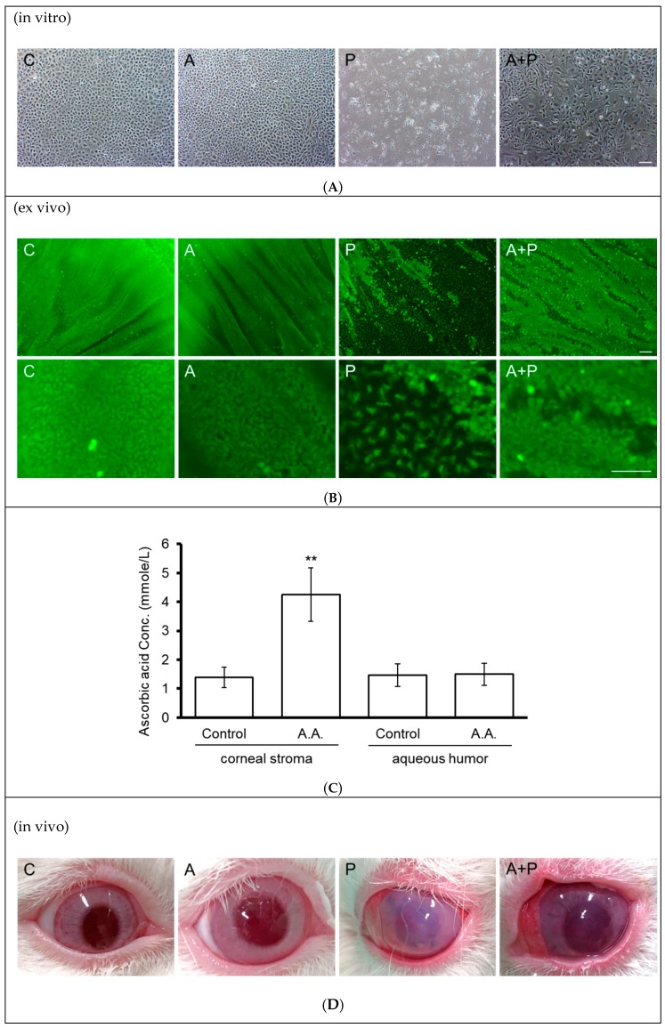
Figure 5.
Effect of topical ascorbic acid on oxidative stress-induced corneal endothelial damage in a rabbit model. (A) Primary rabbit corneal endothelial cells (in vitro) were cultivated in medium with (A and A + P groups) or without (C and P groups) addition of 1 mM of ascorbic acid for two days, followed by addition of paraquat (0.2 mM) in the paraquat-treated groups (P and A + P groups) for five days. Sloughing of corneal endothelial cells was observed under phase-contrast microscopy. (B) Rabbit corneal tissue specimens (ex vivo) were cultivated in medium with (A and A + P groups) or without 1 mM of ascorbic acid for two days, followed by the addition of paraquat (25 mM) to the P and A + P groups for 15 min. Two days later, the sloughing of corneal endothelial cells was observed using Calcein-AM stain. (C) Rabbit corneas (in vivo) received an application of ascorbic acid (284 mmol/L in BSS solution) or BSS alone for two days (three times per day). After diffusion, ascorbic acid concentrations in the corneal stroma and anterior chambers were examined using the FRASC assay. (n = 3, ** p < 0.01). (D) Rabbit corneas (in vivo) received an application of ascorbic acid (284 mmol/L in BSS solution) or BSS alone for two days (three times per day), followed by intracameral injection of 25 mM paraquat (diluted in BSS) or BSS alone for 15 min. Corneal transparency was assessed using external eye photography on day two. (The scale bar represents 100 µm).

