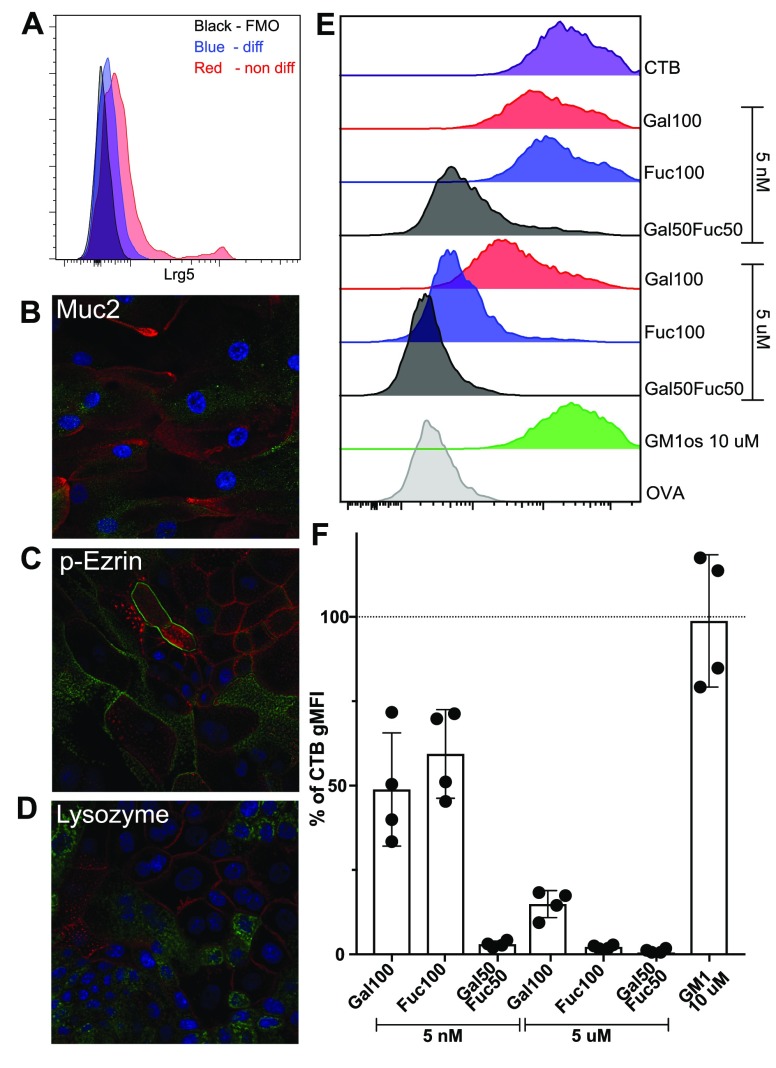
Figure 4.
Enteroid characterization and functional evaluation of the polymer block. (A) Enteroid cells were evaluated using flow cytometry for the presence of Lrg5+ stem cells. (B–D) Enteroid cells were cultured on a transwell insert and differentiated into a non-stem-cell state for 5 days. For all panels, the DAPI stain is blue and the enterocyte marker phalloidin stain is red (for cell visualization). Markers were used to identify different cell types (green) such as goblet cells (B), mature enterocytes (C), and Paneth cells (D). (E) One representative histogram out of two independent experiments of polymer and the GM1-os block of CTB binding to enteroid cells (flow cytometry). Full gating can be seen in Figure S7. One representative analysis out of four donors. (F) Bar graph showing the % of CTB gMFI on cells from four different donors after preincubating CTB with GM1-os or polymers. Error bars are SD.

