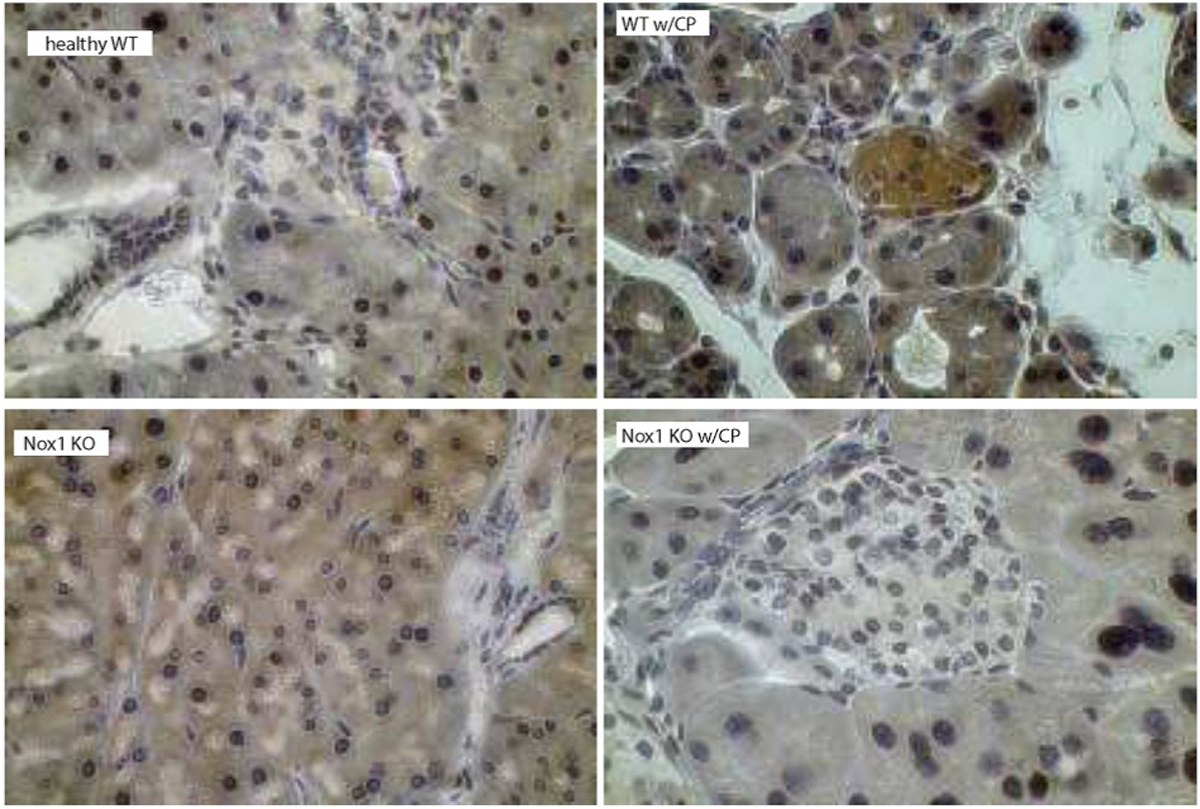
Fig. 4C. Nox1 induces phosphorylation of AKT in a mouse model of CP.

p-AKT was visualized using DAB as a chromogen (color: brown). The staining displayed a cytoplasmic localization of p-AKT in pancreatic acini and islets of Langerhans from WT mice with CP, but not from Nox1 KO mice with CP. Total magnification: 400X.
