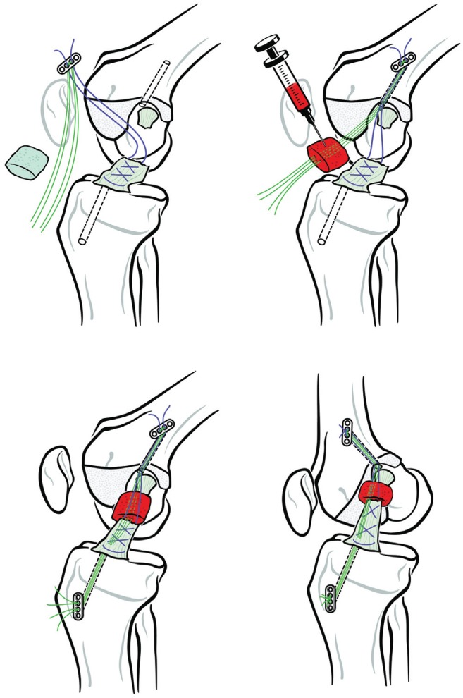
Figure 1.

Schematic of the technique used to place the BEAR implant. Upper left panel: A suture (purple) is placed through the tibial stump via a whipstitch and secured with 2 free sutures (green) to an extracortical button. Upper right panel: After a cortical button carrying free sutures (green) is passed up through the femoral tunnel, the BEAR implant is loaded onto them and soaked with up to 10 mL of autologous blood. Lower left panel: The free suture ends (green) at the tibial end of the BEAR implant (which was positioned between the 2 ends of the torn ACL) are passed through the tibial tunnel to be tied over a second extracortical button. Lower right panel: The sutures and extracortical buttons are secured. ACL, anterior cruciate ligament; BEAR, bridge-enhanced ACL repair.
