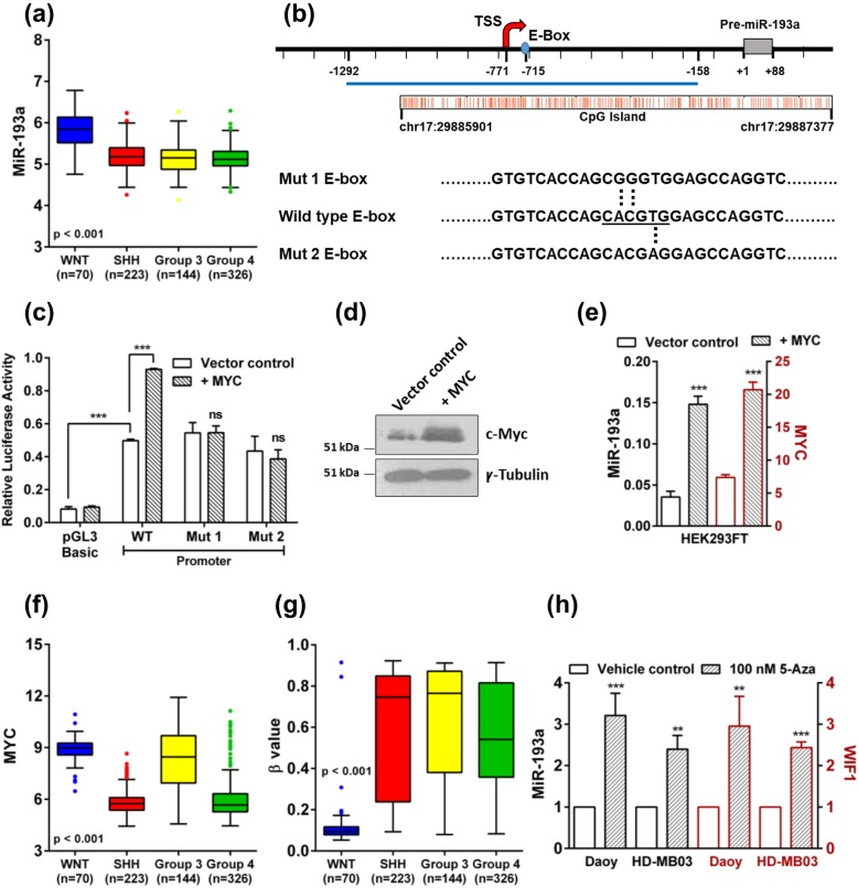
Fig. 1.
Expression of miR-193a, MYC, and the methylation status of the miR-193a promoter region, in the four molecular subgroups of medulloblastomas. Induction of miR-193a expression by MYC, and upon treatment with a DNA methylation inhibitor in medulloblastoma cells. a MiR-193a expression levels in the four molecular subgroups WNT, SHH, Group 3, and Group 4 of 763 medulloblastomas from the MAGIC cohort. b Schematic showing location of the CpG island, E-box, the transcription start site (TSS) relative to the pre-miR-193a start site (+ 1) on chromosome 17 and the mutations introduced in the E-box. c Relative luciferase reporter activity of the miR-193a promoter construct in the presence or absence of MYC and upon the site-directed mutagenesis of the MYC binding site in the miR-193a promoter constructs (Mut 1, Mut 2), upon transient transfection into the HEK293FT cells. d Western blot analysis showing MYC expression in the HEK293FT cells transfected with the MYC expressing plasmid construct. γ-tubulin was used as a loading control. e Induction of miR-193a and MYC expression in the HEK293FT cells transfected with the MYC expressing construct evaluated by real-time RT-PCR analysis. f and g. Expression levels of MYC and the methylation status of a CpG probe (cg22536383) in the miR-193a promoter region, in the four molecular subgroups of medulloblastomas from the MAGIC cohort, respectively. Higher β values indicate higher methylation at the CpG residue. h Fold change in the expression levels of miR-193a and WIF1 in the 5-aza-2′-deoxycytidine treated medulloblastoma cells evaluated by the real-time RT-PCR assay. **, *** and ns indicates p < 0.001, p < 0.0001 and non-significant, respectively

