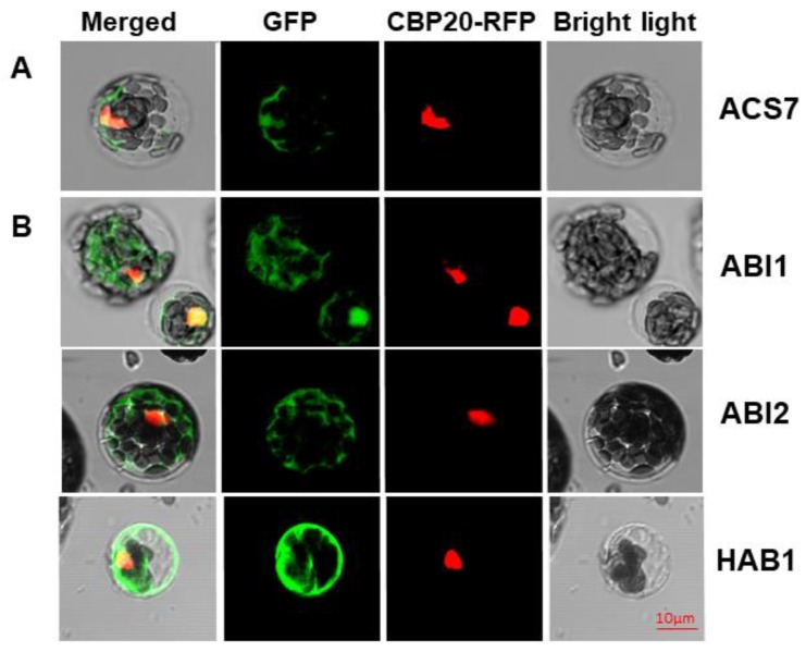
Figure 1.
In planta localization study. Localization of ACS7–GFP (A) and type 2C phosphatases (B) in Arabidopsis thaliana protoplasts, by confocal microscopy. CBP20–RFP was used as nuclear marker. Green fluorescence (ACS7–GFP, ABI1–GFP, ABI2–GFP and HAB1–GFP) and red fluorescence (RFP–CBP20) are merged in the left-hand column with the bright field image; scale bar, 10 µm.

