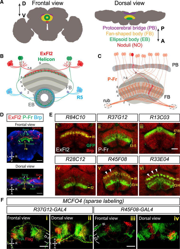
Figure 1. Neuronal Processes Are Organized in Laminae and Columns in the Fan-Shaped Body.
(A) Schematics showing frontal and dorsal views ofthe central complex (CX) in an adult fly brain. The CX is composed of four substructures: the ellipsoid body (EB), the fan-shaped body (FB), a pair of noduli (NO), and the protocerebral bridge (PB). D = dorsal; V = ventral; A = anterior; B = posterior.
B) Schematic showing local sleep circuitry that includes three different types of FB- and EB-projecting neurons. In the FB, ExFl2 neuron axons (red) and Helicon cell dendrites (green) co-stratify in dorsal FB layer 6. In the EB, Helicon cell axons and R5 axons (blue) locally interact with each other in an anterior ring.
(C) Schematic showing small-field P-Fr neuronsthat project processes to the PB, medial FB (mFB) layers 3–5, and an additional brain region called the rubus (rub). Within the PB and the FB, individual P-Fr processes precisely innervate specific dorsal-ventrl columns; P-Fr axons also target the rub. For simplification, the trajectories of P-Fr axons in the diagram do not depict their complete in vivo trajectories.
(D) Two types of FB-projecting neurons are colabeled by using (1) R84C10-lexA to express myristoylated tdTomato (mtdT, red) in about 20 large-field ExFl2 neurons and (2) R37G12-GAL4 to express CD8-GFP (green) in about 30 small-field PBG1–8.s-FℓB[3,4,5.s.b-rub.b (“P-Fr,” green) neurons in an adult fly brain (“s” spine; “b” bouton; “l” layer). Neuropils are revealed here by anti-Brp staining (blue). A frontal view (upper panel) and a dorsal view (lower panel) show that ExFl2 and P-Fr neuron soma are located at posterior-lateral and posterior-medial regions of the brain, respectively. Axons and dendrites project from these soma to the FB and other brain regions.
(E) Multiple types of neurons that target the FB are labeled by different fly lines harboring GAL4 driving expression of mCD8-GFP or lexA driving expression of mtdT (both shown in green in [i]–[vi]). AntiBrp staining (red) labels neuropil structures in adult fly brains. Single optical sections show examples of ExFl2, P-Fr and other types of small-field FB neurons that serve as markers for labeling single, or multiple, FB layers. Small-field FB neurons project their process to confined regions within each layer and form vertical columns orthogonal to FB laminae. These columns are not easily seen via Brp staining, but they can be observed in certain fly lines that label different types of small-field neurons, including R45F08 and R33E04, which label axons in layer 6 (arrowheads).
(F) Subsets of P-Fr neurons (R37G12, [i]–[ii]) and another group of small-field neurons (R45F08, [iii]–[iv]) are sparsely labeled in red and/or green by the multi-color flip-out (MCFO) technique, which illuminates restricted localization of small-field neuron processes in certain FB laminae and columns.
Scale bars represent 50 μm in (D) and 20 μm in (E) and (F).

