Figure 5.

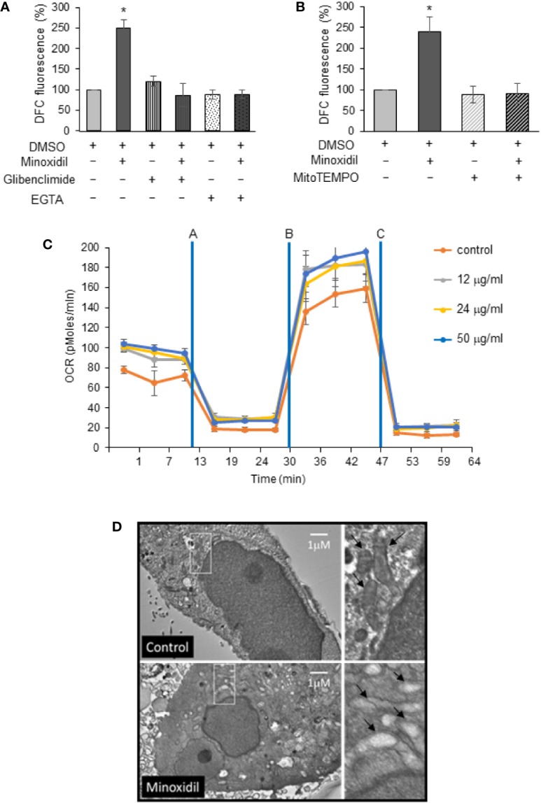
Minoxidil disrupts mitochondria function. (A) Effect of minoxidil alone (50 µg/ml) or with the Kir6.2/SUR blocker glibenclimide (10 µg/ml) or the Ca2+ ion chelator EGTA (2.4 µg/ml) or (B) with MitoTEMPO (5 µg/ml L) compared to control (DMSO) on cellular ROS formation in human-derived OVCAR-8 (DCFH-DA to 2′,7′-dichlorofluorescein DCF, Thermo Fisher Sci; Fluorescence was analyzed in a plate reader (PHERAstar FS, BMG LABTECH) with excitation at 485 nm and emission at 520 nm). Data is expressed as mean ± SEM; *p < 0.001. (C) Kinetic Oxygen Consumption Rate (OCR) of OVCAR-8 cells to minoxidil at 0, 12, 24, or 50 µg/ml. Cells were plated at 25,000/well in XF24 V7 culture plates. The assay medium was the substrate-free base medium. n=3; (Assay design, data analysis, and file management were performed with Agilent Seahorse Wave Desktop software) (D) Representative electron microscopy micrographs showing mitochondria in OVCAR-8 cells treated with minoxidil (50 µg/ml; 24 h). The disruptive effect of the drug is shown as lack of cristae in the mitochondria of treated cells (white box enlarged in right panel).
