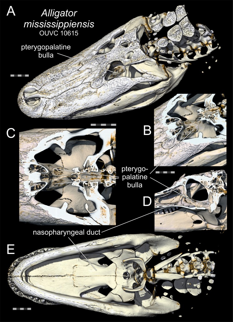
Figure 3. Pterygopalatine bullae of Alligator.
Pterygopalatine bullae of Alligator mississippiensis (OUVC 10615) based on volume renders of computed tomographic data of a fleshy head. (A) Dorsolateral oblique view of the full skull showing the pterygopalatine bulla as seen through the orbit. (B) Dorsolateral oblique view of the pterygopalatine bulla enlarged with the skull roof digitally removed to reveal both bullae. (C) Same presentation as in (B) but in dorsal view and enlarged. (D) Medial (internal) view of the right side of a parasagittaly sectioned head, showing the pterygopalatine bulla emerging as a dorsal dilation of the nasopharyngeal duct. (E) Ventral view of the full skull showing that the pterygopalatine bulla is not visible in ventral view. All scale bars equal 5 cm. (A) and (E) are at the same scale, as are (B) and (D), whereas (C) has its own scale.

