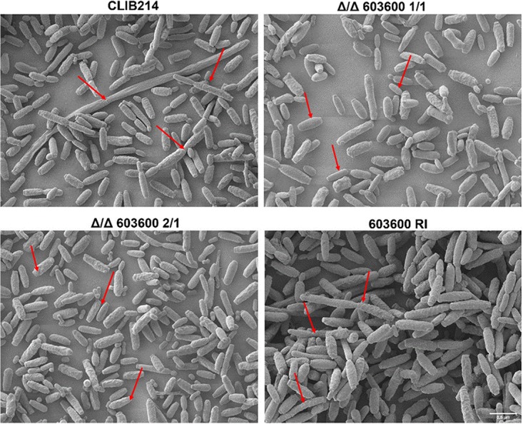
FIG 5.

Scanning electron microscopic images of the wild-type and CPAR2_603600 mutant strains. All strains were visualized using SEM after growth in Spider medium at 37°C for 24 h. Arrows indicate greater numbers of elongated pseudohyphal cells in wild-type and reintegrant strains than in the null mutants, where most of the yeast cells were smaller.
