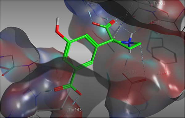
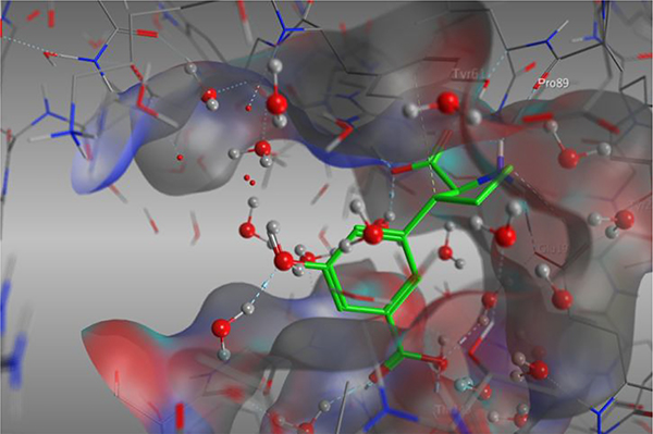
Figure 1.
A) X-ray crystal structure of 5a (green) which holds a 5’-OH substituent, in the LBD of GluA2 (PDB: 4YMA).38 Receptor surface is displayed in grey. B) X-ray crystal structure of 5a (green) in GluA2 (PDB: 4YMA). Protein surface is colored in grey. The 5’-position directs towards cave-shaped space with a depth of 10–12Å.


