
Figure 3.
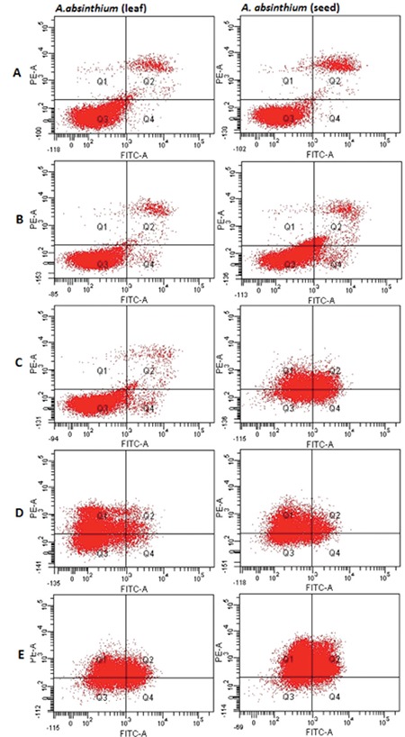
Flow cytometric analysis of apoptosis. A-549 cells were treated with leaf or seed extracts of A. absinthium at various concentrations. Results showing cells in necrosis (Q1), late apoptosis (Q2), live cells (Q3) and early apoptosis (Q4), for control (a), 0.25 mg/mL (b), 0.5 (c), 2 mg/mL (d), 4 mg/mL (e)