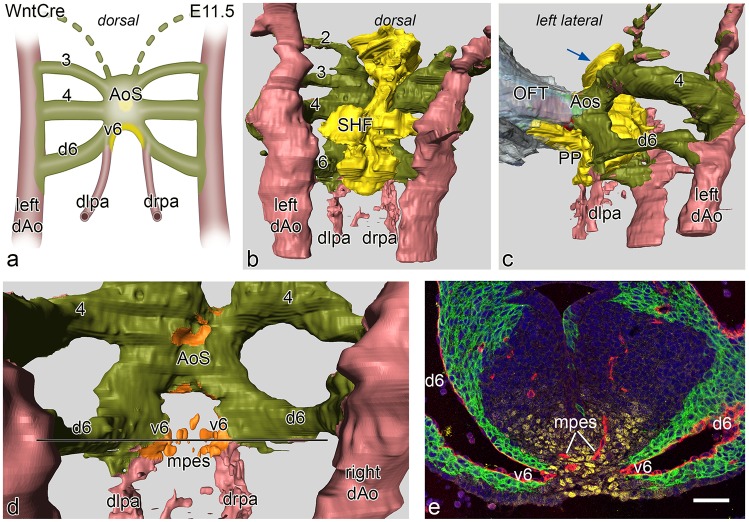
Fig 3.
a. Schematic representation of a dorsal view of vascular connections in an E11.5 WntCre mouse embryo. In this stage PAA 3, 4 and 6 are complete. The distal left (dlpa) and right (drpa) pulmonary arteries merge sideways with PAA 6 creating a ventral (v6) and a dorsal (d6) segment of the latter. The d6 is completely encircled by neural crest cells (green) while the v6 has a lateral layer of neural crest and a medial layer of second heart field (SHF: yellow). The v6 forms a short proximal pulmonary artery. Fig 3b. dorsal view of a reconstruction at this stage with now complete 3rd,4th and 6th arches that are lined by neural crest (olive green). The SHF (yellow) forms the mid-line mesenchyme. The dlpa and drpa (pink) are not covered by either NCC or SHF. Fig 3c. left lateral view showing the connection of the outflow tract (OFT) myocardium (grey) with the short aortic sac (AoS). The SHF mass has a short extension anteriorly towards the future aortic orifice (blue arrow). More prominent at this stage is the left sided SHF extension that runs underneath the v6 and along the future pulmonary wall of the AoS. This is the so-called pulmonary push (PP). Fig 3d. dorsal view after removal of the SHF, the double lining of the relatively short v6 (identical to a proximal part of a pulmonary artery (see also a.) is depicted with a NCC (olive green) and a SHF reflected coverage (orange) Fig 3e. section of the embryo at the level indicated (black line) in d. The right and left v6 (endothelial cells red) are in part lined by NCCs (green) and Nkx2.5 positive SHF (yellow). In the SHF midline mass (yellow) the endothelial plexus (red) of the mid-pharyngeal endothelial strand (mpes) is visible. Magnification: bar 100 μm.

