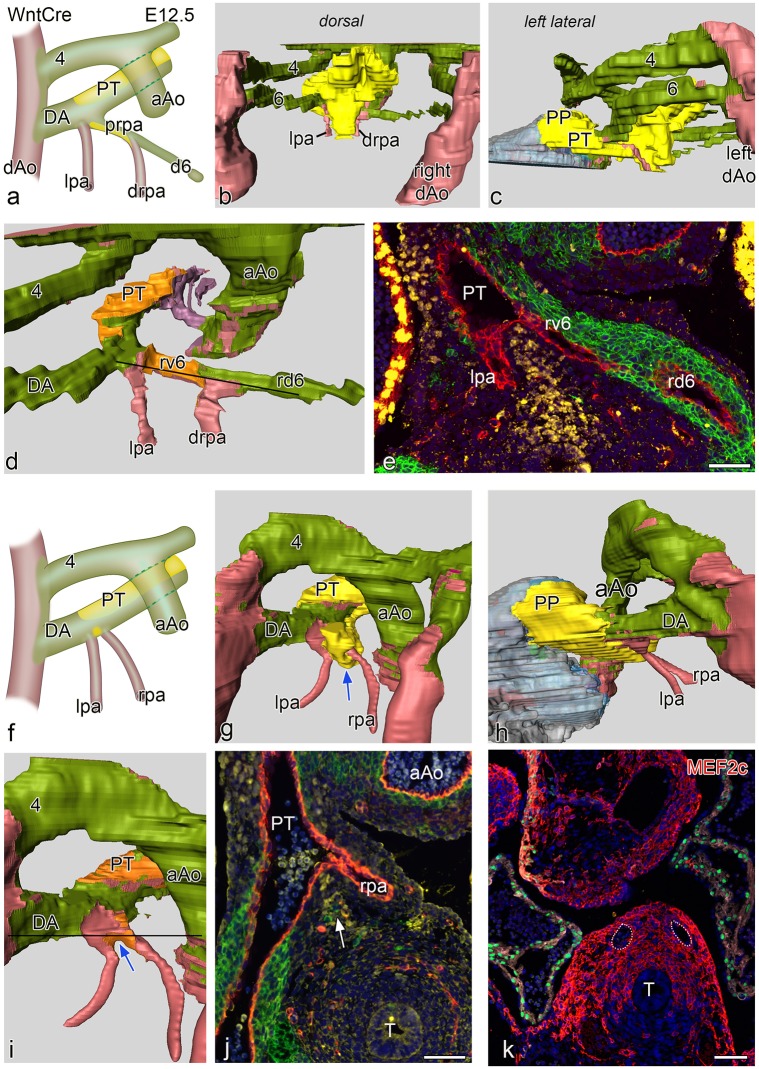
Fig 4.
a and f schematic representations of two E 12.5 WntCre embryos. Embryo a. is slightly less far developed as compared to f. In both embryos the ascending aorta (aAo) and the pulmonary trunk (PT) have now been separated. Fig 4b-4d shows reconstructions of the younger embryo with a complete 4th and 6th PAA. Fig 4c shows the dorsal midline SHF mass (yellow) partly covering the neural crest (green) lined PAA’s. Fig 4c is a left-sided view showing the position of the SHF mass (yellow) which has an extension (pulmonary push population: PP) covering the major part of the lumen of the PT towards the myocardial outflow tract (grey), running underneath the 6th PAA to pulmonary artery connection. Fig 4d. dorsal view after removal of the SHF mass (lumen coverage area: orange). On the left side the dorsal 6th PAA, now referred to as ductus arteriosus (DA) is completely surrounded by neural crest cells, There is no indication of a ventral 6th PAA. The left pulmonary artery (lpa: red) abuts independently on the PT On the right side the situation is less well developed. The dorsal segment of the right 6th PAA (rd6) is regressing and completely surrounded by NCC. The ventral segment of the 6th PAA (rv6) is still present with a lateral wall of NCCs and a medial wall of SHF. The distal part of the right pulmonary artery (drpa) enters side-ways into this right-sided 6th PAA. Thus the proximal rpa is at this stage formed by the rv6. Fig 4e. section at the level indicated in d (black line) in which it can be seen that the right v6 (rv6: also proximal part of the rpa) has both a lining of SHF (yellow) and NCCs (green). Fig 4f in the more developed embryo the originally distal parts of the lpa and rpa, embedded in Mef2c positive mesoderm (Fig 4k) are not covered by NCC and SHF. They enter the PT independent of the DA. Fig 4g-4i are reconstructions showing similar dorsal views as b-d. Level of section j. is indicated by a black line in i. The SHF derived flow divider is still seen between the lpa and rpa (blue arrows in g and i and white arrow in j). The right d6 has regressed completely. There is on both sides no indication anymore of the v6 segments. k section of a E12.5 Mef2cCre embryo showing that the distal parts of the lpa and rpa (white dotted vessels) are situated within Mef2c positive splanchnic mesoderm that is not stained by Nkx2.5 (green). T: trachea Magnification sections e,j,k bars100 μm.

